
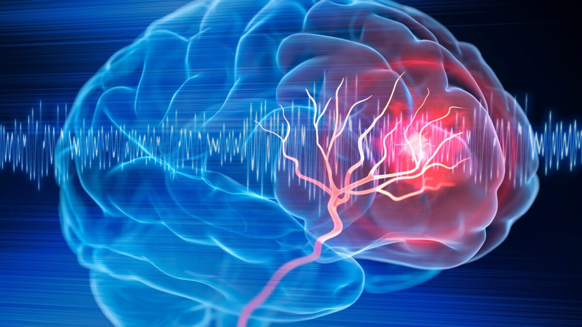
We reviewed the latest technological innovations in radiology and the use of medical imaging and diagnostic testing for traumatic brain injury (TBI) patients. Artificial intelligence usage is mounting with new models making their way into cutting-edge healthcare institutes and medical research centers.
One of the recent developments, credited to neurosurgeons at the UPMC and data scientists at the University of Pittsburgh School of Medicine, involves the use of automated AI models to use brain scans and machine learning algorithms to evaluate traumatic brain injury-sustaining patients.
The development, as of 2022, is a significant step towards implementing artificial intelligence in the field of radiology to enhance medical imaging and diagnostic testing shares David Douglas, assistant professor and staff physician at Stanford University.
He informed me that implementing machine learning could improve patient outcomes and the delivery of vital life-saving healthcare for TBI patients. Based on this development, machine learning-powered AI algorithms can automate brain scans and use relevant clinical data about the patient to assess and predict patient outcomes.
For now, the machine learning model is under further development to enhance its capabilities for quick and accurate predictions about patient survival and recovery. Douglas mentions that TBI is one of the leading causes of disability and death in American children and young adults.
According to the statistics reported by the Centers for Disease Control and Prevention (CDC), nearly 1.5 million people in America sustain a mild to severe traumatic brain injury, resulting in the hospitalization of an estimated 230,000 people.
The report further mentions that nearly 80,000 to 90,000 individuals sustain long-term chronic effects, such as disability. Meanwhile, an alarming 50,000 patients with severe TBI are victims of fatal outcomes. Douglas emphasized the research and development, cost actualization, standardization, and implementation of machine learning models to aid in TBI patient recovery and survival.
With machine learning, clinical radiologists can predict patient outcomes and determine the survival or recovery rate quickly and efficiently. Since AI models have already enabled automated report generation for CT and MRI scans, radiologists can focus on creating individual treatment and healthcare plans for patients.
Most severe TBI patients are admitted to hospitals in critical conditions, prompting neurosurgeons and neurologists to offer immediate surgical or interventional care. This requires quick and efficient medical imaging, which due to the D3D imaging technique has become more detailed and comprehensive.
Douglas developed the Depth-3-Dimensional (D3D) model to increase the sophistication and comprehensibility of CT and MRI scan reports. It allows for accurate spatial depth awareness for greater precision in surgical procedures.
Combined with machine learning, the AI models can leverage medical imaging techniques such as D3D to improve image-taking procedures and provide narrative reports to aid radiologists in providing adequate healthcare.
The current development and scientific studies promise a strong future for AI-powered tools in clinical decision-making. With the advent of artificial intelligence in the medical sector, there is better hope for successful diagnosis and treatment of severe TBI patients admitted in emergency rooms across the US shared Douglas.