
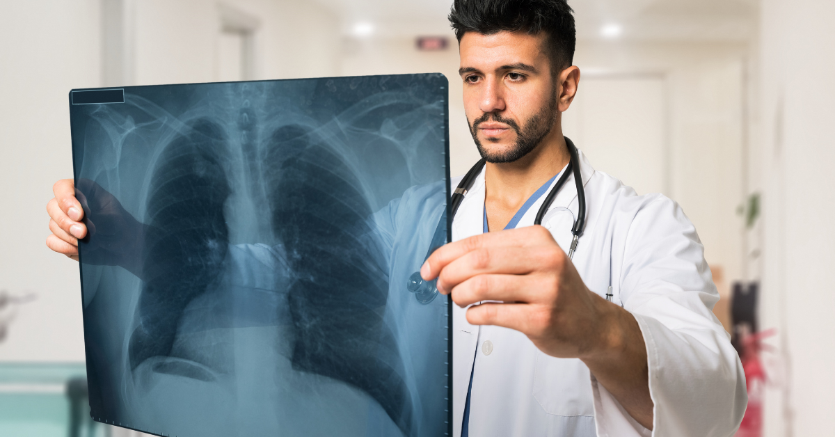
Bronchial artery embolization (BAE) is a method for identifying bronchial routes (supply routes in your lung) using X-ray images. This permits the specialist to find the bronchial corridor which is draining and causing your hemoptysis (hacking up of blood).
Blood vessels and arteries don’t appear on a typical chest X-beam. To see the bronchial corridors a unique color is infused into the course, by means of the crotch, through a fine plastic cylinder called a catheter. X-beams are then taken promptly thereafter, giving us itemized pictures (photos) of the corridors and veins in your lungs. Whenever we have found the bronchial vein that is dying, small particles (the size of grains of sand) are infused to cluster the vessel and stop the death.
Why do You Need a Bronchial Artery Embolization?
Your primary care physician has referred you for a bronchial artery embolization in light of the fact that you are hacking up blood (hemoptysis). The point of a pulmonary artery embolization is to stop or control these manifestations.
Haemoptysis generally happens because of dying, mostly from the bronchial veins (90%) and furthermore from the respiratory supply routes (5%). Draining is regularly more noteworthy when it comes from the bronchial veins in view of the great tension in these courses. Therefore even minor draining from the bronchial veins will in general bring about blood loss.
Irritation of the aviation routes in patients with Cystic Fibrosis or non-CF bronchiectasis may prompt hemoptysis. This irritation causes growth of the bronchial corridors and new vessel development. These amplified vessels and profoundly harmed lung tissue increase the shot of dying particularly during a chest disease. A lung specialist usually recommends embolization.
Before the Procedure
Assuming you are having an arranged methodology you will be approached to go to the Procedure Unit with the goal that we can set you up. At times you will be approached to report directly to X-beam.
You might be seen by one of the physiotherapists who will evaluate your chest. In the event that there is any emissions present that are undermining your breathing, then, at that point, you will be given some delicate physiotherapy to help clear these before the strategy. Assuming you are still effectively draining then you won’t be given physiotherapy.
You will be approached to put on a clinic outfit. The methodology is helped out through the huge course (the femoral vein) in the crotch thus we might request that you shave the skin around the space. The attendant can do this for you assuming you can’t do it without anyone else’s help. Bronchial artery bleeding can be identified through this procedure.
You will have a needle placed into a vein in your arm with the goal that the radiologist can give you a narcotic to assist you with unwinding or to give you pain relievers during the methodology.
A medical assistant will accompany you to the X-beam Department.
Assuming that you have any hypersensitivities, you should let the radiologist know. In the event that you have recently responded to intravenous difference medium (the color utilized for kidney X-beams and CT examinations), you should likewise educate the radiologist concerning this.
Bronchial Artery Embolization Procedure
You are supposed to lie on the X-beam table, for the most part, level on your back. You might have a checking gadget joined to your chest and finger, and might be given oxygen through little cylinders in your nose.
The radiologist will keep everything as sterile as could be expected and will wear a performance center outfit and working gloves. The skin close to the place of inclusion, generally the crotch, will be cleaned with antiseptic, and afterward, the remainder of your body will be covered with a performance center towel.
The skin and more profound tissue over the course will be desensitized with nearby sedatives, and afterward, a needle will be embedded into the conduit. When the radiologist is convinced that this is in the right position, an aide wire is put through the needle, and into the vein. Then, at that point, the needle is taken out permitting a fine plastic cylinder called a catheter to be set over the wire and into the course.
The radiologist utilizes the X-beam gear to ensure that the catheter and the wire are moved into the right position. The color is then infused through the catheter and X-beams are taken. We will request that you pause your breathing while every X-beam is taken.
The radiologist will then, at that point, infuse little particles into the draining vein, which stops the dying.
Toward the end, the radiologist will eliminate the catheter and a solitary line will be utilized to close the passage site.
After the Procedure
Toward the finish of the methodology, the medical caretaker will apply strain to the injury, normally in your crotch, for around 10 minutes after the catheter is eliminated.
You will then, at that point in time, be taken to your ward on a bed where you will rest for 4 hours. You can not get up to go to the toilet during these 4 hours (the medical caretaker can present to you a chamber pot or urinal). For the main hour, you will be elevated level with one cushion.
To try not to drain from the crotch site, it is significant that you don’t twist your leg during this time. The medical attendants on the ward will check your fringe and foot beats just as your blood strain to identify any limitation in the bloodstream to the lower appendages brought about by the methodology performed at a lung specialist.
They will likewise check your crotch site to ensure there is no draining from it. The crotch site will be covered with a mortar that might be eliminated within 24 hours. The fastener used to close the passage site will break down.
You might eat and drink. Drink a lot of liquid as this will assist with flushing the differentiation color through your kidneys.